Pelvis
What determines the fact that a running amateur who runs 10 km in 50 minutes moves in a completely different way than a professional athlete who runs the same distance in less than 27 minutes? It is to a large extent one’s running form, which heavily relies on the position of the pelvis.
Increasing number of people becomes interested in running form and looks for information regarding its propensities, as well as the instructions on developmental stages of movement. The process of arriving at the correct movement is difficult to follow due to the fact that the real-life examples of the pro athletes cannot serve as immediate models. The movement of a proficient runner is impossible to imitate on the go, as it constitutes a process based on certain instinctive and subconscious perceptual mechanisms and neural conduction. Consequently, the conclusion is that the correct running form is impossible to acquire during running itself. Moreover, based on numerous observations and learning attempts, it seems that running is not the easiest form of movement. If it was so, everyone would excel at running form during the very first training.
Training monitoring is increasingly available to runners. The possibilities of taking measurements on one’s own provided by different electronic equipment and the amount of data for training analysis exceeds one’s practical needs. Unfortunately, despite the fact that there are some applications with virtual coaches allowing for choosing training load on the basis of the established goal, there are no applications enabling any analysis of movement or running form. Running industry seems disinterested in launching such applications, focusing on new concepts and systems of feet support. From the commercial point of view, the rest of the body of a runner insignificant. The technical aspects of running are usually left out in amateur sports, due to the fact that they require cooperation and involvement of someone who can register and evaluate one’s movement.
Pelvis matters
The aim of this article is to draw attention to a proposal of running form evaluation based on the position of the pelvis during running. As a relatively new theory, it shouldn’t be taken as the decisive factor in evaluation, but rather as a hint. The theory is backed up by wide experience derived from other sports like martial arts, gymnastics, as well as dancing in which the effectiveness of the movement is associated with the correct pelvis position.
The easiest way to show the correct position of the pelvis is to present a specific example. The simplified animation below shows the rotary motion of the pelvis.
There are a lot of expressions used to describe the position of the pelvis. The clockwise rotation and the moment of the maximal angle in this rotation then can be described as for example, lift, upward inclination or pelvis leveling. There is also a definition of pushing the pelvis forward, although later in the article I will describe it as an incorrect movement.
The return movement, also known as the backward pelvis rotation (a counter-clockwise movement) and the maximal inclination of the pelvis in this rotation can be labeled as a retracted, forward-bent or downward-bent pelvis.
How do the muscles work while running?
Pelvis constitutes the center of the body and the source of effective movement. One cannot perform any effective movement without the pelvis. It applies specifically to such a complex movement as running. The strongest muscles of our bodies are attached to the pelvis, primarily the hip flexors and glutes. Those muscles work properly when the pelvis position is correct. Below there is a selection of the hip line muscles with their descriptions and functions
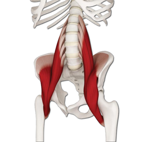
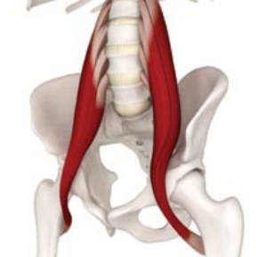
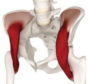
Iliopsoas muscle (top picture) consists of the psoas major muscle (middle picture), the psoas minor muscle (not marked), and the iliacus muscle (bottom picture). While running, the iliopsoas muscle, as its name suggests, plays the role of the main flexor of the hip joint and it raises the thigh bone during leg transfer from the front to the back pendulum. The rectus femoris muscle (a part of quadriceps) and the sartorius muscle also play the role of the hip joint flexors. However, those two muscles are not as strong and effective as the iliopsoas muscle.
 Psoas major muscle
 is attached to the last dorsal vertebra and four lumbar vertebrae at the top. It is not attached directly to the pelvis, but it works together with the iliacus muscle through their common tendon attached to the thigh bone’s lesser trochanter. These long, parallel muscle fibers allow high motion range of the thigh bone.
Iliacus muscle covers the iliac fossa. Its large physiological cross-section, as well as its fan-like arrangement of the muscle fibers allow to produce substantial force. The iliacus muscle moves the thigh bone to which is attached from below (at the lesser trochanter).
Iliopsoas muscle belongs to the group of posture muscles. It is characterized by the increased tone while resting, the ability to sustain long work, high-strain endurance and quick recovery after the effort. Unfortunately, it has a tendency to shortening and contraction, usually due to many hours of sitting which is nowadays a huge part of everyday routine. When lower limbs are not moving, for example while standing, a shortened psoas major muscle pulls the lumbar curve forward, perpetuating the lordosis and possibly causing backache. On the other hand, shortened iliacus muscle can push the pelvis forward and downward. Consequently, a person with a dysfunction of the iliopsoas muscles is not able to lift the pelvis. Instead, such a person pushes it forward and downward to even a greater extent which distorts running biomechanics. In this position any shifts of the body weight between the thigh bones’ heads and pelvis are less effective. The pelvis itself becomes unstable, tilts from side to side and dissipates energy which results in the shortening of the float phase and the suppression of the stride’s elasticity. Moreover, it influences certain geometrical correlations between parts of limbs’ biomechanical chain.
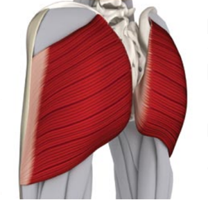
Hip joint extensors are antagonist muscles of the iliopsoas muscle. The main and the strongest hip joint extensor is the gluteus maximus muscle (top picture). The remaining hip joint extensors are the semimembranosus muscle and the semitendinosus muscle.
All dysfunctions like a contracture, myasthenia or arthopy of the muscle in stagnation influence the position of the pelvis and the motion range of the limbs, resulting in the ineffective work of the whole system. Restoring the correct muscle balance and the stability of the pelvis requires medical consultation and posture correction exercises – firstly by relaxing and then strengthening particular muscles. Effective rehabilitation requires time and includes much more complex relations between the muscles and their antagonists than examples presented above.
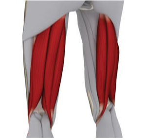
It is very hard to change the pelvis position while running. The pelvis is not as controllable (by one’s will) as for example the foot (one can 'decide’ to land or the heel or the midfoot). It is an important issue and it can suggest that change of the running form is not related to the position of the foot. The weight of the foot is relatively small in comparison to the pelvis and all of the organs inside the pelvis. The foot is too light to play any altering role in the running form. The pelvis is the major player here and landing on the midfoot is only an effect of the correct pelvis position.
Runners who learn to land on the midfoot very often push the foot into the ground while landing. This action is accompanied by a distinctive shuffling sound and the stiffening of the lower leg. Any attempts to eliminate this effect by controlling the foot or changing the length of the stride are not effective in the long term.
Many questions arise concerning the importance of the heel in the landing phase. Referring to the previously mentioned suggestions about the role of the pelvis in generating the movement, shuffling and the role of the heel in the support phase are closely related to the pelvis position. If the pelvis is perfectly balanced and doesn’t have any dysfunctions resulting from some extensive tensions (in other words: any muscle imbalance), then the landing is more fluent and less straining for the upper leg, and the heel does not take part in moving the body’s weight. A correct pelvis position makes it even harder to shuffle or touch the ground with one’s heel.
Elastic recoil
The correct position of the pelvis is closely related to the effect of the elastic recoil. An in-depth description of this phenomena is available in the article devoted to running rhythm (see:
Rhythm). Elastic recoil depends on the elasticity of one’s body in order to move. Such a potential can be fully realized only after a correction to the position of the pelvis has been made (suppression of the elasticity of every step needs to be minimized). A retracted pelvis is the main suppressor of this effect. A lifted pelvis provides a visualization and an impression of a very dynamic take-off, and consequently, relaxation of the moving limbs.
The images below show the examples of a retracted and a lifted pelvis. The animations were created using the frontal plane. The choice of the orientation is not random because the frontal plane enables the analysis of the elastic recoil effect and the position of the hips.
How do the best run?
For the purpose of the comparison I selected two elite athletes. The animations show Moses Mosop, who is a leading long-distance runner with marathon PB of 2:03:06 (Boston 2011) and 10km PB of 26:49, and Jan Frodeno, a triathlete and the Olympic champion from Bejing with 10 km PB of 29:20.
I’ve chosen those two athletes, because there is limited access to the footage which would show runners in comfortable training conditions without the influence of stress caused by competition
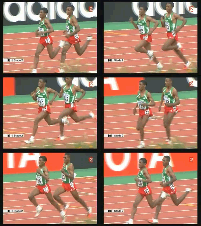
The analysis of Moses Mosop’s running enables observation of the correct position of the hips. It is characterized by a leveled, horizontal hip line during the critical moment of the support phase. His running in itself is very dynamic and seems relaxed. Dynamics of the take-off stems from the maximal usage of elasticity. Consequently, the impression of relaxation stems from the cohesion of the figure moving in a harmonious, coordinated rhythm.
In the case of Jan Frodeno’s, there is a distinctive image of “sitting” on the supporting leg, which is indicated by tilted hip line compensated by tilted shoulder line during the support phase. It is a very distinctive feature that can signal the retracted pelvis. Moreover, it is accompanied by a significant suppression of the elasticity and his running looks like hard, muscular and not very dynamic work.
A retracted pelvis, and consequently, the movement of the whole biomechanical chain can affect the process of cumulative fatigue but in Mosop’s case it is incremental or incidental (hip line leveled to such extent in the frontal plane is a very rare phenomenon that can be observed among the fastest runners in the world). When it comes to the retracted pelvis of Frodeno, it is constant and independent from the pace and the level of fatigue.
The example of Jan Frodeno displays a very typical case of “sitting” on the supporting leg during the support phase. This is a flaw from point of view of motion effectiveness, but it can be observed among majority of runners, both professionals and amateurs. It is a phenomenon that exists independently from one’s physique and performance level.
One can accurately evaluate the position of the pelvis by watching tilts of the hip and shoulder lines in the frontal plane. It may serve as a proposal of a criterion for running form evaluation that can be conducted without any special equipment. The only thing needed is a well positioned camera to capture the support phase.
Summary
While searching for the reasons behind the incorrect position of the pelvis during running one reaches a saddening conclusion, which is that the entire problem stems from the modern lifestyle. Its effects are especially visible during mass running events. Long hours of sitting and slouching contribute to perpetuation of the retracted position of the pelvis. In order to reverse these harmful effects, stretching is necessary. In this respect, special attention should be payed to iliopsoas muscle (which stabilizes the pelvis) and its antagonists. However, it does not constitute a ultimate solution to the problem of the correct position during running.
The running equipment market is continuously proposing another state-of-the-art designs of running shoes which are supposed to improve the dynamics of the take-off. Such innovative models aim at pushing the limits of human body. It seems that a world-class runner displaying a correct pelvic position would not need any additional dynamics-improving equipment. On the other hand, for the majority of amateur runners even the most innovative shoes would not overcome the obstacle posed by a retracted pelvis.
References:
Ray Long The key muscles of Hatha Yoga

 Psoas major muscle is attached to the last dorsal vertebra and four lumbar vertebrae at the top. It is not attached directly to the pelvis, but it works together with the iliacus muscle through their common tendon attached to the thigh bone’s lesser trochanter. These long, parallel muscle fibers allow high motion range of the thigh bone.

